Thoracentesis
What is thoracentesis?
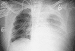
Critically ill or injured patients may accumulate fluid around one or both lungs due to a number of different disease processes. The fluid around the lung is called a pleural effusion. The procedure that the doctors perform to sample or drain the pleural effusion is called thoracentesis.
How is thoracentesis performed?
The procedure is usually performed at the patient's bedside in the ICU. The doctor first uses numbing medicine (local anesthetic) to numb an area of skin in between the ribs and then a small needle and/or plastic tube (catheter) is placed into the pleural effusion. Some or all of the fluid is removed and sent to the laboratory for testing. The tests may reveal why the fluid developed and help guide how best to treat it. Usually the catheter and the needle are removed after the thoracentesis and a chest X-ray is taken to assess the lung.
Does thoracentesis hurt?
Thoracentesis is associated with minimal discomfort. Even this is reduced by use of a local anesthetic.
Are there any potential complications associated with thoracentesis?
Potential complications associated with thoracentesis include bleeding, infection, and lung collapse.
Chest X-ray showing large left pleural effusion (right side of picture)
